White Line Disease in Horses
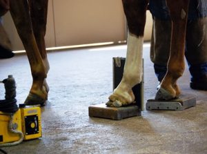
Photo: Erica Larson, News Editor
Radiographs should be taken to locate precisely
the extent of the disease’s progression and to verify
the angle of the coffin bone.
When bacteria and fungus creep into the living layers in the horse’s foot, they can cause extensive damage known as white line disease.
The many layers of the hoof make it a living, growing, highly functional, powerful structure capable of supporting the weight of the entire horse.
Unfortunately, this construction also puts the horse at risk of hoof infections within the layers. One of the most common of these is white line disease.
Despite its name, white line disease does not actually affect the true “white line” of the hoof, which can be seen on the sole where it joins the hoof wall and appears more yellow than white, according to Stephen E. O’Grady, DVM, BVSc, MRCVS, a farrier and veterinarian specializing in equine podiatry at Northern Virginia Equine in Marshall. White line disease occurs when bacterial or fungal infections creep into the inner nonpigmented space within the inner hoof wall’s stratum medium layer. The infection slowly eats away the tissue, which turns into a chalky powder that spills out when scraped with a hoof pick.
“You can hear that hollowness behind the hoof wall wherever there’s separation just by tapping lightly with a farrier hammer,” O’Grady says.
Without treatment, white line disease continues to travel up through the layer, sometimes causing lameness, and it will eventually reach (but not involve) the coronary band. At that point the laminae, which attach the hoof wall to the coffin bone, can separate and cause the coffin bone to rotate downward.
“If rotation occurs, the prognosis of white line disease-induced mechanical laminitis is very bad,” O’Grady says. “It’s critical to address the problem before it progresses that far, because the good news is that it can be successfully treated.”
How is it Treated?
Every treatment plan should begin with collaboration between a veterinarian and an informed farrier, according to Klaus Mäurer, a master blacksmith, nationally accredited farrier instructor in Germany, and education chair at the European Federation of Farriers Association (EFFA).
As white line disease is often misunderstood, he notes it’s best to work with professionals who have had continuous training with knowledgeable experts.
Radiographs should be taken to locate precisely the extent of the disease’s progression and to verify the angle of the coffin bone, Mäurer says. The farrier and veterinarian should then design an immediate shoeing strategy to support the hoof during treatment.
Treating white line disease requires resection, or removal, of the hoof wall wherever the disease has spread. “The fungi and bacteria responsible for white line disease are anaerobic (they grow best without oxygen), so what we have to do is get that area completely exposed to the oxygen in the air,” says Denis Leveillard, international farrier trainer, vice president of the French farriers’ association, and a former president of the EFFA.
A support shoe should be applied immediately before resection to prevent unequal pressure on the remaining hoof wall. “I like to use a bar shoe to help the sole handle the horse’s weight and his force when he takes a step,” Mäurer says. Rubber or synthetic soles can hold antibiotics and antifungal creams in contact with the white line, and silicone can be used for sole support. Nails should be as small and few as possible, he says, and they can be substituted with special glues if the hoof wall is unable to hold nails.
Resection of the nonsensitive outer hoof capsule can be carried out painlessly with normal farrier tools in most situations, O’Grady says. However, the farrier should not reach blood or live tissue.
Once the wall has been cut away, the newly exposed layer should be debrided (dead tissue removed), or thoroughly cleaned, to get rid of all bacteria, fungi, and powdery residue.
Cracks can be filled with appropriate fillers, Mäurer says. But under no circumstances should the resected area be covered with acrylics or other chemicals, as maintaining exposure for treatment is vital in order to cure white line disease.
What is the Healing Process?
It takes approximately one year for a hoof to grow out completely from the coronary band to the end of the toe, our sources note. Thus, healing time can be calculated according to how high the wall was cut to treat white line disease.
Special hoof care is necessary until the wall has grown back completely. “It’s essential that the horse owner and the stable personnel understand how important it is to have a special cleaning management plan for that horse,” Mäurer says.
Debriding should be performed at least twice a month, and the hoof should be kept dry, according to O’Grady.
If the resection does not go higher than halfway up the hoof wall, and if sufficient support has been provided by appropriate shoeing, you can continue to ride the horse and give him light exercise during healing, O’Grady says.
How Does it Happen?
No one really knows why horses develop white line disease. The disease is common worldwide in mares, stallions, and geldings of all breeds, ages, and conditions.
Even so, it does seem that trauma or poor conformation could open the door–or more literally the hoof–to disease. Opportunistic bacteria and fungi find their way up into the hoof through separations and cause a secondary infection, which then becomes white line disease. Humid conditions might make white line disease more likely, or they can make a case worse.
White line disease tends to reappear in horses that have had it previously, according to O’Grady. So if you’ve had to treat one hoof, make sure you have all hooves on that horse checked regularly for any signs of disease.
TakeHome Message
For all horses, have well-trained, qualified farriers maintain regular trimming and hoof care. They can shape the hooves to help prevent separations and recognize white line disease in its early stages, leading to quick, minimized intervention and rapid recovery.
ABOUT THE AUTHOR
Christa Lesté-Lasserre, MA
Christa Lesté-Lasserre is a freelance writer based in France. A native of Dallas, Texas, Lesté-Lasserre grew up riding Quarter Horses, Appaloosas, and Shetland Ponies. She holds a master’s degree in English, specializing in creative writing, from the University of Mississippi in Oxford and earned a bachelor’s in journalism and
creative writing with a minor in sciences from Baylor University in Waco, Texas. She currently keeps her two Trakehners at home near Paris. Follow Lesté-Lasserre on Twitter