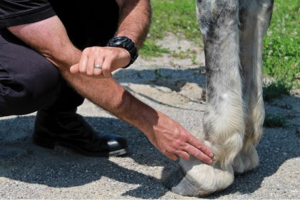
Bone chips can be a proper pain in the joint; learn where and why they happen and when they need to be removed.
Posted by Stacey Oke, DVM, MSc | Oct 7, 2015 | Article, Forelimb, Lower Limb, Muscle and Joint Problems, Musculoskeletal System, Surgical Techniques, Thoroughbred
Small, but mighty, bone chips can be a proper pain in the joint
It wasn’t so long ago that mention of a broken-this or a shattered-that meant a horse’s demise. But with time, research, and improved imaging technology, veterinarians have determined that some fractures aren’t so terrible. One such example is a classic chip fracture.
Chips, short for bone chips, are technically osteochondral fragments—pieces of cartilage-covered bone (“osteo” for bone and “chondral” for cartilage) that have “chipped” off, often times into a joint.
“Osteochondral ‘chip’ fragments are common in athletic horses, especially racehorses,” says Robert J. Hunt, DVM, MSc, Dipl. AVCS, surgeon at Hagyard Equine Medical Institute, in Lexington, Kentucky. “With proper management, chips do not have to be either the career- or life-threatening problem that other types of fractures are.”
Bones chip in a variety of ways and for a number of reasons, and veterinarians sometimes have their work cut out for them in finding these miniscule insults among the protuberances, eminences, and cuboidal bones that comprise the complex equine skeleton. In this article we’ll discuss these tiny chips that can cause big pain, and, of course, what to do about them.
Why Does Bone Chip?
Horse’s bones might seem like rods of steel, but even steel has its breaking point, as we learned from the Titanic. Hunt explains that there are three ways bones can fracture. In the first scenario, the bone structure fails due to momentary loading beyond its strength. This can occur due to acute trauma from getting kicked, striking a firm object, or falling down, among other misadventures. Other chips occur because the bone has weakened due to infection, neoplasia (tumors), osteoporosis (reduced bone mass), or other bone disease, making it unable to withstand normal amounts of loading. This is called a pathological fracture. Finally, an accumulation of microdamage and an imbalance between bone remodeling and resorption (bone breakdown, which is a normal part of bone turnover) can lead to a fatigue fracture. Specifically, microdamage accumulation causes excessive resorption, making the bones prone to fracture. “Fatigue fracture is the most common cause of chip fractures within joints of racehorses,” says Hunt. [pullquote source=”Dr. Robert J. Hunt”]The carpus and fetlock are far more likely to be victims of chip fractures than other joints. [/pullquote] Chips can range in size from flakes to pebbles or even slabs of bone. As with any fracture, clinical signs include sudden onset of heat, pain, fluid accumulation within the affected joint, and lameness.Where the Chips Lie
Theoretically, chips can fracture off the edge of any bone, but veterinarians see them most commonly in specific joints. “The carpus (knee) and fetlock are far more likely to be victims of chip fractures than other joints because of focal cyclical loading (repeated weight-bearing) on a relatively small surface area together with a high range of motion,” says Hunt. “Those factors allow stress concentration and weakening of the underlying bone. Eventually, the straw breaks the camel’s back, and a chip will displace from parent bone. It is generally the integrity of the underlying bone and the surrounding cartilage or other soft tissues that determine the prognosis for return to function.” Within those vulnerable joints are “preferred” locations for chipping, he adds. Some of the top locales for bone chips include:- The proximal (top) or distal (bottom) aspect of the radial carpal or intermediate carpal bones;
- The third carpal bone (these can be simple chips or full slab fractures, in which the fracture extends completely from one side of the bone to the other);
- The distal radius (both medial and lateral—or inner and outer—aspects);
- Any one of the eminences (small, protruding areas of bone) of the proximal aspect of the first phalanx (long pastern bone) in the fetlock; and
- The sesamoid bones, either of the fetlock or the navicular.
Scrutinizing Chips
Veterinarians have a number of ways to diagnose even the smallest of fragments. After conducting a physical and lameness examination, radiography (taking X rays) is usually the first tool practitioners reach for when they suspect a horse has a chip fracture. Veterinarians recommend taking a series of standard views of the affected joint from a variety of angles to best determine chip size, shape, and exact location. Sometimes chips are difficult to find, and it can take several X ray views to locate them. Other times fragments don’t displace completely from the bone, making fracture lines difficult to visualize. Despite its wide recognition as a soft-tissue diagnostic tool, ultrasonography is useful for diagnosing chip fractures if chips occur in lower limb joints, particularly around joint margins, says Roger Smith, MA, VetMB, PhD, DEO, Dipl. ECVS, MRCVS, a professor of equine orthopedics at the Royal Veterinary College, in the United Kingdom. Veterinarians can also use MRI to diagnose bone chips, but this modality is less widely available, and MRI equipment is more expensive than X ray units. Similarly, CT, nuclear scintigraphy (bone scanning), and thermography are additional but far less commonly used options for detecting equine osteochondral chip fractures.Surgery vs. Conservative Care
Treatment approach all boils down to whether the horse can live with a chip or if he’d be better off without. The answer to that problem is not always obvious. “Information about the use of the horse, location of the chip, presence of more than one chip in a joint, more than one affected joint, size or shape of the chip, and whether there is evidence of degenerative joint disease all help veterinarians decide whether surgery is the best option or not,” says Hunt. Consider some of the following cases:- Most horses (except for elite show jumpers, due to the repeat concussion on landing) can tolerate fractures in the top (radiocarpal) joint of the knee;
- Chip fractures in the lower joint of the knee will remain a constant problem for all horses except those involved in only very light work, such as infrequent trail riding;
- Pleasure horses with well-rounded chip fractures (a sign that the chip has been present for some time) and no evidence of active degenerative joint disease often do not require surgery; and
- Chip fractures at the proximal and dorsal (front) aspect of the first phalanx can be found in sound horses and do not necessarily cause lameness. That said, surgeons might need to remove those same chip fractures in some performance horses that are not living up to their owner’s expectations.
 Maps courtesy of Centers for Disease Control and Prevention; Tick photos courtesy of Mat Pound/USDA/Bugwood.org and Wikimedia Commons
Learn the latest on diseases horses can get from ticks and why they continue to frustrate veterinarians and researchers
If the sight of a tick makes your skin crawl—even if it’s not crawling on your skin— you’re not alone. That feeling is founded on more than a natural aversion to arachnids; diseases transmitted by ticks can pose a real health threat. With Centers for Disease Control and Prevention (CDC) maps outlining tick ranges throughout the majority of the United States, it’s important we brush up on our understanding of tick-borne diseases. In this article we’ll take a look at the three that pose the biggest risk to horses: Lyme disease, anaplasmosis, and piroplasmosis.
Lyme Disease
Horse owners living in areas of the country heavily infested with Ixodes scapularis, commonly known as blacklegged ticks (also referred to as deer ticks or bear ticks), know these parasites are more than a nuisance. In these regions contracting Lyme disease from infected ticks is entirely possible for horses and humans alike.
Lyme disease is a very difficult disease to prevent, diagnose, and treat in horses, says Linda Mittel, MSPH, DVM, senior extension associate at Cornell University’s Animal Health Diagnostic Center, in Ithaca, New York. Horses contract Lyme disease when the spirochete (a type of bacterium) Borrelia burgdorferi is transmitted through the bite of an infected tick.
“Signs of Lyme disease might not appear until 6 weeks after exposure”
Diagnosis Here’s why diagnosing Lyme disease is tricky: Not all infected horses exhibit clinical signs, and the signs themselves are often confounding, meaning they can point to any number of other diseases. Even if you know an infected tick bit your horse (You can actually test it!), signs of Lyme disease might not appear for up to six weeks after exposure. In short, nothing is straightforward with this disease. An infected tick might or might not transmit the bacteria to its host, and an infected animal might or might not exhibit signs of disease.
So while Lyme disease symptoms are not necessarily cut-and-dried, they can include low-grade fever, muscle tenderness, muscle wasting, weight loss, a stiff or uncoordinated gait, swollen joints, “shifting” lameness in various joints, lethargy, hyperesthesia (sensitivity to sound and touch), and uveitis (simply, inflammation of the vascular layer of the eye). While in private practice in the Northeast, Mittel often saw non-neurologic clinical signs of Lyme disease that included unexplained weight loss, changes in attitude and behavior, uveitis, and hypersensitivity. Some signs might arise from the bacterial infection itself, while others (e.g., uveitis) result from the body’s immune response to it. Lyme neuroborreliosis (NB), a rare condition, occurs when B. burgdorferi infects the horse’s nervous system.
Maps courtesy of Centers for Disease Control and Prevention; Tick photos courtesy of Mat Pound/USDA/Bugwood.org and Wikimedia Commons
Learn the latest on diseases horses can get from ticks and why they continue to frustrate veterinarians and researchers
If the sight of a tick makes your skin crawl—even if it’s not crawling on your skin— you’re not alone. That feeling is founded on more than a natural aversion to arachnids; diseases transmitted by ticks can pose a real health threat. With Centers for Disease Control and Prevention (CDC) maps outlining tick ranges throughout the majority of the United States, it’s important we brush up on our understanding of tick-borne diseases. In this article we’ll take a look at the three that pose the biggest risk to horses: Lyme disease, anaplasmosis, and piroplasmosis.
Lyme Disease
Horse owners living in areas of the country heavily infested with Ixodes scapularis, commonly known as blacklegged ticks (also referred to as deer ticks or bear ticks), know these parasites are more than a nuisance. In these regions contracting Lyme disease from infected ticks is entirely possible for horses and humans alike.
Lyme disease is a very difficult disease to prevent, diagnose, and treat in horses, says Linda Mittel, MSPH, DVM, senior extension associate at Cornell University’s Animal Health Diagnostic Center, in Ithaca, New York. Horses contract Lyme disease when the spirochete (a type of bacterium) Borrelia burgdorferi is transmitted through the bite of an infected tick.
“Signs of Lyme disease might not appear until 6 weeks after exposure”
Diagnosis Here’s why diagnosing Lyme disease is tricky: Not all infected horses exhibit clinical signs, and the signs themselves are often confounding, meaning they can point to any number of other diseases. Even if you know an infected tick bit your horse (You can actually test it!), signs of Lyme disease might not appear for up to six weeks after exposure. In short, nothing is straightforward with this disease. An infected tick might or might not transmit the bacteria to its host, and an infected animal might or might not exhibit signs of disease.
So while Lyme disease symptoms are not necessarily cut-and-dried, they can include low-grade fever, muscle tenderness, muscle wasting, weight loss, a stiff or uncoordinated gait, swollen joints, “shifting” lameness in various joints, lethargy, hyperesthesia (sensitivity to sound and touch), and uveitis (simply, inflammation of the vascular layer of the eye). While in private practice in the Northeast, Mittel often saw non-neurologic clinical signs of Lyme disease that included unexplained weight loss, changes in attitude and behavior, uveitis, and hypersensitivity. Some signs might arise from the bacterial infection itself, while others (e.g., uveitis) result from the body’s immune response to it. Lyme neuroborreliosis (NB), a rare condition, occurs when B. burgdorferi infects the horse’s nervous system.
 While Lyme disease symptoms are not necessarily cut-and-dried, they can include low-grade fever, muscle tenderness, muscle wasting, weight loss, a stiff or uncoordinated gait, swollen joints, “shifting” lameness in various joints, lethargy, hyperesthesia (sensitivity to sound and touch), and uveitis (simply, inflammation of the vascular layer of the eye).
Photo: iStock
Researchers have made strides in Lyme disease diagnostic testing, but there’s still a long way to go. The newest test for B. burgdorferiantibodies (the immune system produces antibodies to fight antigens, or foreign substances, it detects) in the blood is the equine Lyme Multiplex assay developed by the Animal Health Diagnostic Center. This screening identifies three antigen proteins:
While Lyme disease symptoms are not necessarily cut-and-dried, they can include low-grade fever, muscle tenderness, muscle wasting, weight loss, a stiff or uncoordinated gait, swollen joints, “shifting” lameness in various joints, lethargy, hyperesthesia (sensitivity to sound and touch), and uveitis (simply, inflammation of the vascular layer of the eye).
Photo: iStock
Researchers have made strides in Lyme disease diagnostic testing, but there’s still a long way to go. The newest test for B. burgdorferiantibodies (the immune system produces antibodies to fight antigens, or foreign substances, it detects) in the blood is the equine Lyme Multiplex assay developed by the Animal Health Diagnostic Center. This screening identifies three antigen proteins:
 ABOUT THE AUTHOR
 Horses developing laminitis might shift the weight off their feet twice as much as they do normally.
Photo: iStock
Your horse’s best chance of overcoming this hoof disease might lie in your ability to catch it early
It’s a painful condition that veterinarians, farriers, and horse owners have been racking their brains about for decades. Laminitis—the separation or failure of laminae, which connect the hoof wall to the coffin bone within—can cause permanent structural changes in a horse’s foot, leading to repeated bouts of disease and lasting lameness. In severe cases the pedal (coffin) bone in the hoof rotates downward, potentially even puncturing the sole and prompting the decision to euthanize. But get this: Watchful handlers can actually detect signs of laminitis in its early stages and intervene before the condition becomes debilitating.
“Everyone talks about laminitis being a lameness issue, but we know that horses start to get damage at a microscopic level before they show any lameness,” says Andrew van Eps, BVSc, PhD, MACVSc, Dipl. ACVIM, senior lecturer and specialist in equine medicine at The University of Queensland Equine Hospital, in Gatton, Australia.
Therefore, keeping an eye out for minute changes in your horse’s health is key to maximizing his likelihood of recovery, says Tom Ryan, FWCF, a researcher and farrier based in Bedfordshire, U.K. “You have to be proactively thinking ahead,” he says.
To help you catch this devastating hoof disease while your horse still has a chance to avoid suffering its consequences, our sources have helped us come up with a list of 10 early warning signs. Regardless of the type of case (-supporting-limb, systemic inflammatory response syndrome, or endocrine disease-related), these red flags could indicate laminitis is setting in—even before you see any signs of lameness. So alert your veterinarian as soon as possible if you detect one or more of the following:
Horses developing laminitis might shift the weight off their feet twice as much as they do normally.
Photo: iStock
Your horse’s best chance of overcoming this hoof disease might lie in your ability to catch it early
It’s a painful condition that veterinarians, farriers, and horse owners have been racking their brains about for decades. Laminitis—the separation or failure of laminae, which connect the hoof wall to the coffin bone within—can cause permanent structural changes in a horse’s foot, leading to repeated bouts of disease and lasting lameness. In severe cases the pedal (coffin) bone in the hoof rotates downward, potentially even puncturing the sole and prompting the decision to euthanize. But get this: Watchful handlers can actually detect signs of laminitis in its early stages and intervene before the condition becomes debilitating.
“Everyone talks about laminitis being a lameness issue, but we know that horses start to get damage at a microscopic level before they show any lameness,” says Andrew van Eps, BVSc, PhD, MACVSc, Dipl. ACVIM, senior lecturer and specialist in equine medicine at The University of Queensland Equine Hospital, in Gatton, Australia.
Therefore, keeping an eye out for minute changes in your horse’s health is key to maximizing his likelihood of recovery, says Tom Ryan, FWCF, a researcher and farrier based in Bedfordshire, U.K. “You have to be proactively thinking ahead,” he says.
To help you catch this devastating hoof disease while your horse still has a chance to avoid suffering its consequences, our sources have helped us come up with a list of 10 early warning signs. Regardless of the type of case (-supporting-limb, systemic inflammatory response syndrome, or endocrine disease-related), these red flags could indicate laminitis is setting in—even before you see any signs of lameness. So alert your veterinarian as soon as possible if you detect one or more of the following: