Health & Care Tips
Read the Latest News

Fighting White Line Disease in Horses

fighting-white-line-disease

Your vet and farrier should work together to debride affected tissue and kill the fungi.

Photo: Erica Larson, News Editor

The late famed farrier Burney Chapman long ago stressed the importance of treating white line disease (WLD) aggressively and considering the overall picture rather than just the pathology (disease or damage). The fungi involved in white line disease attack the stratum corneum’s (the outermost layer of the epidermis) intermediate layer—called the stratum medium—and digest the membrane responsible for securing the hoof wall to the sensitive areas deeper in the center of the hoof. The end result is that air and debris, instead of cartilage, line and separate the living tissue layers. Tapping the hoof wall in these cases creates a hollow sound, which is why some call WLD hollow hoof syndrome.

This disease’s complex management and treatment aspects are probably best narrowed to three components: hoof care, diet, and environment. Ignoring any one of these can cause an otherwise good plan to fail.

Hoof care

A veterinarian and farrier team is best suited to address individual WLD cases, so we will cover only general principles here. First, the affected area must be opened and dried out with a topical product such as chlorine dioxide or Gentian violet. The degree of damage will dictate the amount of hoof wall debridement required, and care must be exercised not to damage healthy tissue or create more damage to the wall than is prudent—the horse still must bear weight comfortably. Treatment is aimed at killing fungi or fungal spores rather than trying to burn or harden sensitive tissue, as the latter actually slows the overall healing process. 

Second, the vet-farrier team should provide coffin bone support either by casting or shoeing and remove flares, seedy toe, and lamellar wedges (abnormal horn structure that develops within the lamellar region of the foot) to establish a proper weight-bearing structure for the horse. Finally—a step that’s often overlooked—your farrier should trim and shoe the horse to achieve a heel-first landing and proper weight bearing over the solar surface of the foot. 

Diet

We know many horses that suffer from WLD are insulin resistant or have equine metabolic syndrome, and we know forage type, quality, and quantity directly influence insulin levels. So we must consider feed types and sources for affected horses. Insulin resistance, for instance, can cause blood vessels at the extremities to contract, potentially compromising blood flow to the foot’s tissues, so we’d focus on a low-calorie/low-carb diet. 

Environment

Horses with poor blood circulation in their feet require adequate exercise to stimulate blood flow, so turnout and pasture time are critical to overall success. Stalling rather than turning out in a paddock environment may be the single biggest factor compounding pathogen growth and damage associated with white line disease. Horses moving even in small paddocks are much better equipped to battle opportunistic hoof pathogens than those living stationary in a stall environment. 

Hoof form and function has developed into an entire field of study and, as we gain knowledge pertaining to current practices, we have begun to rethink our current animal husbandry guidelines or practices. Researchers are examining how bedding types and footing (e.g., pea gravel vs. sand) affect foot growth and function. Results from one study showed that changes in confinement and footing materials were superior to current white line disease and laminitis treatment techniques. 

There is much more to white line disease than hoof wall separation or an ideal environment for fungi to become pathogenic. We need to realize that fungi are opportunistic and will continue to cause problems until we debride and treat the affected areas. While it’s still open for some debate, many researchers generally believe WLD is tied to the way we house and care for our horses. Volumes are being written on this subject, and the answers we seek will affect the future care and husbandry of our horses.

White Line Disease in Horses

  White line disease  

Photo: Erica Larson, News Editor

Radiographs should be taken to locate precisely the extent of the disease’s progression and to verify the angle of the coffin bone.
   
  When bacteria and fungus creep into the living layers in the horse’s foot, they can cause extensive damage known as white line disease.
      The many layers of the hoof make it a living, growing, highly functional, powerful structure capable of supporting the weight of the entire horse.    
Unfortunately, this construction also puts the horse at risk of hoof infections within the layers. One of the most common of these is white line disease.
   
Despite its name, white line disease does not actually affect the true “white line” of the hoof, which can be seen on the sole where it joins the hoof wall and appears more yellow than white, according to Stephen E. O’Grady, DVM, BVSc, MRCVS, a farrier and veterinarian specializing in equine podiatry at Northern Virginia Equine in Marshall. White line disease occurs when bacterial or fungal infections creep into the inner nonpigmented space within the inner hoof wall’s stratum medium layer. The infection slowly eats away the tissue, which turns into a chalky powder that spills out when scraped with a hoof pick.
   
“You can hear that hollowness behind the hoof wall wherever there’s separation just by tapping lightly with a farrier hammer,” O’Grady says.
   
Without treatment, white line disease continues to travel up through the layer, sometimes causing lameness, and it will eventually reach (but not involve) the coronary band. At that point the laminae, which attach the hoof wall to the coffin bone, can separate and cause the coffin bone to rotate downward.
   
“If rotation occurs, the prognosis of white line disease-induced mechanical laminitis is very bad,” O’Grady says. “It’s critical to address the problem before it progresses that far, because the good news is that it can be successfully treated.”
   
How is it Treated?
   
Every treatment plan should begin with collaboration between a veterinarian and an informed farrier, according to Klaus Mäurer, a master blacksmith, nationally accredited farrier instructor in Germany, and education chair at the European Federation of Farriers Association (EFFA).
   
As white line disease is often misunderstood, he notes it’s best to work with professionals who have had continuous training with knowledgeable experts.
   
Radiographs should be taken to locate precisely the extent of the disease’s progression and to verify the angle of the coffin bone, Mäurer says. The farrier and veterinarian should then design an immediate shoeing strategy to support the hoof during treatment.
   
Treating white line disease requires resection, or removal, of the hoof wall wherever the disease has spread. “The fungi and bacteria responsible for white line disease are anaerobic (they grow best without oxygen), so what we have to do is get that area completely exposed to the oxygen in the air,” says Denis Leveillard, international farrier trainer, vice president of the French farriers’ association, and a former president of the EFFA.
   
A support shoe should be applied immediately before resection to prevent unequal pressure on the remaining hoof wall. “I like to use a bar shoe to help the sole handle the horse’s weight and his force when he takes a step,” Mäurer says. Rubber or synthetic soles can hold antibiotics and antifungal creams in contact with the white line, and silicone can be used for sole support. Nails should be as small and few as possible, he says, and they can be substituted with special glues if the hoof wall is unable to hold nails.
   
Resection of the nonsensitive outer hoof capsule can be carried out painlessly with normal farrier tools in most situations, O’Grady says. However, the farrier should not reach blood or live tissue.
   
Once the wall has been cut away, the newly exposed layer should be debrided (dead tissue removed), or thoroughly cleaned, to get rid of all bacteria, fungi, and powdery residue.
   
Cracks can be filled with appropriate fillers, Mäurer says. But under no circumstances should the resected area be covered with acrylics or other chemicals, as maintaining exposure for treatment is vital in order to cure white line disease.
   
What is the Healing Process?
   
It takes approximately one year for a hoof to grow out completely from the coronary band to the end of the toe, our sources note. Thus, healing time can be calculated according to how high the wall was cut to treat white line disease.
   
Special hoof care is necessary until the wall has grown back completely. “It’s essential that the horse owner and the stable personnel understand how important it is to have a special cleaning management plan for that horse,” Mäurer says.
   
Debriding should be performed at least twice a month, and the hoof should be kept dry, according to O’Grady.
   
If the resection does not go higher than halfway up the hoof wall, and if sufficient support has been provided by appropriate shoeing, you can continue to ride the horse and give him light exercise during healing, O’Grady says.
   
How Does it Happen?
   
No one really knows why horses develop white line disease. The disease is common worldwide in mares, stallions, and geldings of all breeds, ages, and conditions.
   
Even so, it does seem that trauma or poor conformation could open the door–or more literally the hoof–to disease. Opportunistic bacteria and fungi find their way up into the hoof through separations and cause a secondary infection, which then becomes white line disease. Humid conditions might make white line disease more likely, or they can make a case worse.
   
White line disease tends to reappear in horses that have had it previously, according to O’Grady. So if you’ve had to treat one hoof, make sure you have all hooves on that horse checked regularly for any signs of disease.
   
Take­Home Message
   
For all horses, have well-trained, qualified farriers maintain regular trimming and hoof care. They can shape the hooves to help prevent separations and recognize white line disease in its early stages, leading to quick, minimized intervention and rapid recovery.
   
ABOUT THE AUTHOR
   
Christa Lesté-Lasserre, MA Christa Lesté-Lasserre is a freelance writer based in France. A native of Dallas, Texas, Lesté-Lasserre grew up riding Quarter Horses, Appaloosas, and Shetland Ponies. She holds a master’s degree in English, specializing in creative writing, from the University of Mississippi in Oxford and earned a bachelor’s in journalism and
creative writing with a minor in sciences from Baylor University in Waco, Texas. She currently keeps her two Trakehners at home near Paris. Follow Lesté-Lasserre on Twitter


Hoof Abscesses in Horses

  Hoof Abscess

Photo: The Horse Staff

A hoof abscess is like a whitehead pimple:
   
  That little bubble of pus under the skin can be slightly sore or it can be incredibly painful, and the fastest way to get rid of it is to pop it and let it drain.
   
  This common problem can cause sudden , severe lameness, but it often can be resolved quickly with proper treatment approach.
   
  Yesterday your horse was perfectly sound, but today he won’t touch one foot to the ground. There is no apparent injury or sign of a problem in his leg or hoof other than the sudden, severe lameness. What could have happened?
   
  One strong possibility for the cause of this scenario is a hoof abscess–a localized accumulation of pus within the horse’s hoof.  The good news is that the abscesses can often be resolved quickly and easily with proper veterinary care and leave no lasting damage. Even better, practicing good routine hoof care and management can usually prevent them.
     
As an owner, how do you prevent abscesses and what do you do if your horse gets one? First let’s discuss what they are and what causes them.
       
 
Hoof Zits
   
  The simplest comparison we can make to define a hoof abscess is that it’s like a whitehead pimple. That little bubble of pus under the skin can be slightly sore or it can be incredibly painful.  You might feel soreness in that location well before the pimple shows it’s ugly head, or it might show up overnight in all it’s glory.  And the simplest way to get rid of it is simply to pop it and let it drain; the pain relief is immediate because the pressure has been relieved.
   
This is the same way a hoof abscess causes pain in a horse; it usually starts with a localized, walled-off infection, which the body fights with white blood cells and inflammatory mediators. The buildup of infection, inflammation, and white blood cells expands, causing increasing pressure, particularly because the rigid hoof wall can’t expand to relieve the pressure. When lameness appears and how severe the lameness becomes will vary. Some Horses might never get lame before the abscess ruptures on its own, or lameness might be transient and go unnoticed, especially if the horse is at pasture and not monitored often.
What Causes Abscesses?
Most abscesses begin with bacteria entering interior hoof structures, usually via the sole-wall junction (just inside the hoof wall). Anything that weakens hoof/sole integrity can make it easier for bacteria to invade, and internal hoof injuries (such as bruising) can also result in abscesses. Following is a list of common causes:
     
Environmental Conditions cycling between wet and dry
     
In very dry conditions the hoof dries out and can shrink slightly like a dried-out sponge. This can result in tiny hoof cracks and fissures in the sole-wall junction that can then soften and fill with muck when the weather turns wet, allowing opportunistic bacteria to invade the hoof and cause an abscess.
     
Penetrating Wounds
      Penetrating wounds can occur as a result of a horse stepping on a sharp object such as a nail, rock, or broken glass. “These may cause a perforation of the sole that packs up and seals over, and an abscess results two to four days later as a result of contamination,” says Bruce Lyle, DVM, of the Aubrey Equine Clinic in Aubrey, Texas.  
     
“Close” nails in a recently shod foot
     
Raul Bras, DVM, of Rood & Riddle Equine Hospital in Lexington,Kentucky, explains that a horseshoe nail placed too close to or into the foot’s sensitive inner structures can introduce bacteria that cause an abscess.. Even if the nail is removed right away and didn’t introduce bacteria, it created a pathway into the hoof that can let in bacteria and result in an abscess later. Bras recommends flushing the hole with dilute antiseptic solution, such as Betadine, and wrapping the foot for three to five days (depending on the horse’s turnout situation) to decrease the chances of infection.
     
Ground conditions/bruising
     
Muddy or rocky ground can soften feet and/or cause bruises.  “Some non-penetrated bruises may abscess if bacteria are introduced through a small external insult or from circulating bacteria in the bloodstream, because the area of hemorrhage provides a great medium for bacterial reproduction,” Lyle says.
     
Hot-fitting a shoe on a very thin sole
     
Lyle says if the sole is very thin and a hot shoe is seated on it, thermal injury to the underlying sensitive tissues can sometimes cause a sterile abscess (not caused by infection). Applying exothermic (heat-producing)  hoof  repair materials over raw or partly healed areas can do the same thing
 
   
Poor hoof balance/conformation  
     
For example, hoof wall flares can put additional bending stress on the sole-wall junction and cause cracks that can become contaminated. Also, Lyle says leaving the bars on the foot too long (or leaving any part of the foot longer so it gets more of a beating) can result in localized bruising and abscesses.
     
Management factors   
     
Dirty stalls tend to be wet and contain lots of bacteria that can invade the foot. “Wet conditions are usually the culprit in our area, especially when preceded by dry conditions,” says Lyle.
Bras notes that hoof/wall capsule defects can also make it easier for bacteria to invade, as can digital instability (such as that resulting from severe laminitis) or systemic infections. With the latter, bacteria in the bloodstream get into the foot tissues and “set up shop,” causing an abscess from within.
         

Detecting Abscesses


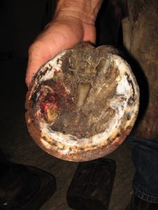
“Clinical signs depend on the severity of the infection; therefore, lameness could vary from mild, minimal lameness progressing to moderate, severe lameness,”  says Bras.  “Other clinical signs might include swelling, heat, draining tracts (pus, often gray or black in color, from the sole.coronary band), increased digital pulse, and evidence of hood injuries (that can introduce bacteria into inner hoof structures, leading to abscesses)”.
 
In severe cases deep within the hoof, the abscess pocket or its effects, such as deteriorating bone, are visible on a radiograph.
“A hoof tester exam applying focal force is often vital to localizing an abscess within the confines of the foot,” says Lyle. “As the pressure increases, so does the pain.”
Also, when trimming the foot one might see a black spot on the sole or sole-wall junction where a crack or puncture is contaminated with muck. This stands out in contrast to the rest of the clean, trimmed sole. This contaminated tract might lead to an abscess (not all contaminated cracks will cause abscesses). Bras notes that most abscesses can be found this way.
       
    Abscess Treatment
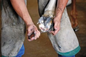
Similar to treating pimples, the basic abscess treatment strategy is to open it and let it drain. Some will even pop on their own, often after traveling up the hoof to the coronary band or heel bulbs where the wall is thinner and easier to break through.
     
When possible, a veterinarian drains an abscess through the sole for two reasons: One, the cack or puncture that can lead to an abscess generally is in the sole, and it can be followed to the abscess. Two, this puts  a hole beneath the abscess so gravity can help pull out the pus. Cleanliness is essential during and after the procedure.
“Treatment requires cleaning the foot, locating the entry wound (if there us one), establishing drainage, softening the hoof capsule via foot soaks and poultices to encourage rupture/drainage, and keeping the foot wrapped and protected from further debris entering causing further infection,” says Bras. “Anti-inflammatory medication and antibiotics may also be given if needed. After drainage is obtained, progressive improvement should be expected on a daily basis.  “I f drainage and lameness continue, perform other diagnostic procedures to determine the true cause.
     
Preventing Abscesses
   
     
  “Good hoof care that leaves adequate sole for protection and develops a snug and uniform sole-wall junction is the best line of prevention,” says Lyle. Good hoof care includes frequent hoof cleaning to remove rocks/mud and routine farrier care to keep the feet balanced and address any problems. “If a horse has thin soles or is prone to bruising…protect them with shoes, etc.,” says Bras. “Keep the feet trimmed so they don’t get wall separations that can lead to white line disease and abscesses. Be proactive; don’s wait for things to happen.”
   
  Take-Home Message
   
Lyle explains, “The most important thing to know about abscesses is to get your lame horse looked at as early in the process as possible by a veterinarian who’s interested in horses. Abscesses generally are straightforward and shouldn’t require extravagant and expensive imaging to diagnose or treat, although exceptions do exist.”
   
 
THE AUTHOR: CHRISTY M. WEST:
   
Christy has a BS in Equine Science from the University of Kentucky, and an MS in Agricultural Journalism from the University of Wisconsin-Madison
Do You Have a Tip or an Idea for a Story? Tell Us About It.