Tick-Borne Disease: Tremendously Tricky in Horses

Tick-Borne Disease: Tremendously Tricky in Horses
  • May 6, 2017
Tick Borne Disease Map Maps courtesy of Centers for Disease Control and Prevention; Tick photos courtesy of Mat Pound/USDA/Bugwood.org and Wikimedia Commons Learn the latest on diseases horses can get from ticks and why they continue to frustrate veterinarians and researchers   If the sight of a tick makes your skin crawl—even if it’s not crawling on your skin— you’re not alone. That feeling is founded on more than a natural aversion to arachnids; diseases transmitted by ticks can pose a real health threat. With Centers for Disease Control and Prevention (CDC) maps outlining tick ranges throughout the majority of the United States, it’s important we brush up on our understanding of tick-borne diseases. In this article we’ll take a look at the three that pose the biggest risk to horses: Lyme disease, anaplasmosis, and piroplasmosis. Lyme Disease Horse owners living in areas of the country heavily infested with Ixodes scapularis, commonly known as blacklegged ticks (also referred to as deer ticks or bear ticks), know these parasites are more than a nuisance. In these regions contracting Lyme disease from infected ticks is entirely possible for horses and humans alike. Lyme disease is a very difficult disease to prevent, diagnose, and treat in horses, says Linda Mittel, MSPH, DVM, senior extension associate at Cornell University’s Animal Health Diagnostic Center, in Ithaca, New York. Horses contract Lyme disease when the spirochete (a type of bacterium) Borrelia burgdorferi is transmitted through the bite of an infected tick. Signs of Lyme disease might not appear until 6 weeks after exposure” Diagnosis Here’s why diagnosing Lyme disease is tricky: Not all infected horses exhibit clinical signs, and the signs themselves are often confounding, meaning they can point to any number of other diseases. Even if you know an infected tick bit your horse (You can actually test it!), signs of Lyme disease might not appear for up to six weeks after exposure. In short, nothing is straightforward with this disease. An infected tick might or might not transmit the bacteria to its host, and an infected animal might or might not exhibit signs of disease. So while Lyme disease symptoms are not necessarily cut-and-dried, they can include low-grade fever, muscle tenderness, muscle wasting, weight loss, a stiff or uncoordinated gait, swollen joints, “shifting” lameness in various joints, lethargy, hyperesthesia (sensitivity to sound and touch), and uveitis (simply, inflammation of the vascular layer of the eye). While in private practice in the Northeast, Mittel often saw non-neurologic clinical signs of Lyme disease that included unexplained weight loss, changes in attitude and behavior, uveitis, and hypersensitivity. Some signs might arise from the bacterial infection itself, while others (e.g., uveitis) result from the body’s immune response to it. Lyme neuroborreliosis (NB), a rare condition, occurs when B. burgdorferi infects the horse’s nervous system. Tick Borne Disease Hors While Lyme disease symptoms are not necessarily cut-and-dried, they can include low-grade fever, muscle tenderness, muscle wasting, weight loss, a stiff or uncoordinated gait, swollen joints, “shifting” lameness in various joints, lethargy, hyperesthesia (sensitivity to sound and touch), and uveitis (simply, inflammation of the vascular layer of the eye). Photo: iStock Researchers have made strides in Lyme disease diagnostic testing, but there’s still a long way to go. The newest test for B. burgdorferiantibodies (the immune system produces antibodies to fight antigens, or foreign substances, it detects) in the blood is the equine Lyme Multiplex assay developed by the Animal Health Diagnostic Center. This screening identifies three antigen proteins:
  • “Outer surface protein A” antibodies, which were previously thought to be found in vaccinated horses, but now the research is undecided;
  • “Outer surface protein C” antibodies in recently infected horses, which are present within three to five weeks of infection, declining by seven to 11 weeks and no longer present by five months; and
  • “Outer surface protein F” antibodies in horses with chronic infection, identifiable from two to three months on.
These results leave room for interpretation. “What is considered positive is a little hard to specifically define,” notes Mittel. “If one of these areas, particularly the outer-surface protein F, is positive, it doesn’t mean it’s causing disease in the horse; it simply means the animal has been exposed. If the animal is showing clinical signs associated with Lyme disease, in many cases the outer-surface protein F will be elevated, but not always. It’s a very difficult disease to diagnose, because we are, at this point in time, diagnosing with an ‘all antibody test’—not testing for the organism (itself) in the body, but for the animal’s response to the organism. Antibodies mean the horse has been exposed to the agent. It’s possible the animal will be ill, but a positive doesn’t necessarily mean the horse needs to be treated.” The good news is that the turnaround on the Multiplex test is less than a day. “This gives you an idea if the animal has been exposed,” says Mittel, allowing the veterinarian to begin treatment immediately if the horse has compatible clinical signs. While the Lyme Multiplex is the exciting new kid on the block, another test provides the most definitive analysis. “The Western blot test is the most comprehensive test for antibodies to B. burgdorferi and enables the diagnostician to determine the presence of antibodies produced to all of the major protein components of the organism, rather than the three proteins targeted by the Multiplex test,” says Sandra Bushmich, DVM, MS, section head at the University of Connecticut’s Veterinary Medical Diagnostic Laboratory, in Storrs. “This can prove especially advantageous when following the course of an infection over time or determining response to antibiotic treatment. Because of its increased complexity, the Western blot test takes a few days to run and requires a laboratory with particular expertise in equine Lyme Western blot testing.” “The Western blot test is the most comprehensive test for antibodies to B. burgdorferi.”
  1. SANDRA BUSHMICH
  In addition to Lyme Multiplex and Western blot diagnostics, other available initial screening tests include the canine IDEXX SNAP; the enzyme-linked immunosorbent assay (ELISA); and the indirect fluorescent antibody tests (IFAT), all of which can detect B. burgdorferi antibodies, indicating exposure to Lyme disease. If you find a tick on your horse and save it for evaluation, your veterinarian can submit it to a reference lab for a polymerase chain reaction (PCR) test. This checks the tick for B. burgdorferi DNA. A positive reading indicates it is a vector (carrier) of Lyme disease. This means the horse might have been exposed to the bacteria, but he won’t necessarily develop the condition, explains Mittel. Labs can even use PCR tests to test for the organism within the horse, including within cerebrospinal fluid, joint fluid, and joint tissue, although Mittel notes this procedure is relatively uncommon. Beyond all these testing options, she emphasizes that the veterinarian’s evaluation of the animal and ruling out conditions that produce similar signs is of primary importance. A licensed vaccine providing protection against B. burgdorferi would be a welcome addition to horse vaccination protocols, but currently none is available. Some veterinarians vaccinate horses using a canine product off-label, but this procedure is not without concerns, and its efficacy is questionable. Mittel explains that if a vaccinated horse does develop Lyme disease symptoms, his vaccination status could confound the equine Lyme Multiplex data. Treatment For treatment Mittel says veterinarians most commonly prescribe oral doxycycline and minocycline, both antibiotics. Veterinarians can also administer intravenous oxytetracycline, which, Mittel explains, like all tetracycline antibiotics, has an additional anti-inflammatory response. “If the horse has another condition, like arthritis, which is causing inflammation, and it’s not clear if lameness is due to Lyme disease or another condition, the horse may appear improved after treatment—you may have a false impression that the horse had Lyme disease, when it’s improving to the side effect of drug,” says Mittel. Or, if the horse stays sound after finishing treatment, Lyme disease might have been making him lame. In tick-infested areas, horse owners should check horses often that have been on pasture or in brushy and wooded areas. Remove ticks immediately. According to the CDC website, transmission of the B. burgdorferi bacterium occurs 36-48 hours after the tick has begun feeding. Anaplasmosis Equine granulocytic anaplasmosis, another tick-borne bacterial disease, caused by Anaplasma phagocytophilum (previously known as Ehrlichia equi, with the condition called equine granulocytic ehrlichiosis), is spread by the bite of infected I. scapularisand I. pacificus (Western blacklegged) ticks and infects the host’s white blood cells. Clinical signs, apparent within one to three weeks of infection, might include high fever, suppressed appetite, edema (fluid swelling), depression, petechial (pinpoint) hemorrhage, and inability to walk normally. While anaplasmosis causes some signs similar to Potomac horse fever (PHF), cases tend to occur from late fall through winter and into early spring, whereas PHF occurs in late summer and early fall. Diagnosis Veterinarians can diagnose anaplasmosis by manually evaluating a blood smear with an in-house complete blood count panel. The organism is detectable in white blood cells, explains Mittel, who notes that practitioners might also employ PCR and/or IFAT. Bushmich describes new developments in anaplasmosis diagnosis: “Recent work in our lab has shown a PCR test on a buffy coat sample (the white blood cell layer of a centrifuged whole blood sample) is more sensitive than a PCR on whole blood or direct microscopic exam for morulae (visible inclusion bodies in the white blood cells). … The buffy coat sample PCR is a sensitive test (it produces very few false negatives); this is a really good test for an acutely ill horse (to differentiate it from from other clinically similar diseases). If the veterinarian is considering anaplasmosis, this test is fast—you can usually get it within a day or two, and it’s very specific,” says Bushmich, meaning it produces very few false positives. “Serological (antibody) tests are better for determining previous exposure or subacute (between acute and chronic), long-term cases.” Treatment “In general, this is a self-limiting disease,” meaning it will resolve on its own, says Mittel, “but it can be critical to some horses. Anaplasma responds well to treatment. The drugs of choice are doxycycline or oxytetracycline, both of which are renal (kidney) toxic, so care must be taken during treatment, as the animal may have become dehydrated over the course of the illness.” As with Lyme disease, there is currently no equine vaccine available. Piroplasmosis Transmission of equine piroplasmosis (EP, also called babesiosis), a blood-borne disease caused by Babesia caballi and/or Theileria equi parasite infection, occurs when a tick feeds on an infected horse and then on an uninfected horse, or when contaminated equipment is shared among horses, such as used dental and tattoo equipment, needles, and surgical instruments. Horses can also acquire the disease from infected blood transfusions and from an infected dam during birth. “What’s concerning is the likely spread of (piroplasmosis) between horses, which we see happening with contaminated equipment or blood doping at nonsanctioned Quarter Horse racetracks. ”
  1. LINDA MITTELL
Currently, the disease is not considered endemic in the United States and has occurred only in isolated outbreaks. Because the USDA has declared EP a reportable disease, veterinarians must notify authorities before collecting diagnostic samples in suspected cases. All horses entering the United States, whether temporarily or permanently, must test EP-negative to be allowed entry. Some exceptions to this procedure have been made in the cases of international events, such as the 2010 World Equestrian Games in Kentucky, where the USDA granted EP-positive horses waivers, allowed them entry to compete, and housed and managed them under special conditions with state/federal oversight. Various ticks have been shown, either naturally or experimentally, to be EP vectors. Chantal Rothschild, DVM, Dipl. ACVIM, of Northwest Equine Veterinary Associates, near Seattle, Washington, says that the tropical horse tick (Dermacentor [Anocentor] nitens) and Southern cattle tick (Rhipicephalus [Boophilus] microplus) are proven -transmitters, as are the Cayenne tick (Amblyomma cajennense) and American dog tick (Dermacentor variabilis). A native of Brazil who did her initial veterinary studies there but her internship and residency in the United States, Rothschild has seen EP from various angles. Warming Temperatures Politics aside, data indicates an ongoing trend toward warming temperatures, which in turn impacts habitats and organisms, say scientists at the Cary Institute of Ecosystem Studies in Millbrook, New York, which has released findings from its research center. “Host-seeking behavior by ticks is inhibited by temperatures below freezing, and tick activity slows dramatically below about 40°F,” says Richard Ostfeld, PhD, a disease ecologist there. “Warmer winter temperatures mean that ticks can be active longer, which probably increases their ability to find hosts. We have found that the larval and nymphal stages (nymphs transmit the vast majority of Lyme disease) become active a few weeks earlier in warmer than in cooler years. The long-term trend has been for ticks to emerge earlier.” Along with increased tick activity, warmer weather means expanded ranges and greater numbers of cases. “There is evidence from several studies that, as the climate warms and growing seasons lengthen, ticks are able to invade areas that were formerly too harsh,” he notes. “This is probably why ticks (and the diseases they transmit) have been steadily expanding in both latitude and altitude. The blacklegged ticks (Ixodes scapularis) that transmit Lyme disease, babesiosis, and anaplasmosis don’t appear to have strong effects on their wild animal hosts. But, the winter tick (Dermacentor albipictus) can cause declines in health of their hosts, which are mainly moose and deer, but also sometimes horses. There is some evidence that warm winter conditions increase winter tick infestations by decreasing body condition of the hosts and possibly increasing tick survival.” NATALIE DEFEE MENDIK Infected horses exhibit signs one to two weeks after exposure. Signs can be mild or acute, ranging from suppressed appetite and weakness to fever, anemia, edema, labored breathing, and jaundice. Mittel says cases can run from subclinical (not showing signs) to fatal. Surviving horses, while no longer sick, can pose a biosecurity problem. “Once infected, horses tend to become carriers, even after drug treatment to resolve the clinical signs. They may be still positive years after infection,” says Rothschild. “And here lie the USDA’s concerns: Carrier horses have no clinical signs and cannot be distinguished from normal healthy horses (without diagnostic testing); however, they still have the potential to transmit the disease via ticks or contaminated equipment. “If a horse is known to be EP-positive, it must be euthanized, exported from the U.S., entered into lifelong quarantine, or enrolled in an ongoing research -treatment program under the oversight of the USDA,” she adds. Diagnosis Currently the C-ELISA blood test is the gold standard for EP diagnostic testing, says Rothschild. “It picks up on horses that have low levels of -infection—ones that may test negative (on the complement fixation test, a blood test used to screen horses entering the country from the 1970s to 2005) when, in fact, they are positive.” When the disease is in a chronic state, notes Rothschild, the horse’s antibody levels might fluctuate, complicating test results. DNA testing is also an option. A USDA–approved laboratory must conduct both these tests. “What’s concerning is the likely spread of this disease between horses, which we see happening with contaminated equipment or blood doping at nonsanctioned Quarter Horse racetracks,” remarks Mittel. “The concern is then if an infected racehorse retires and becomes part of the mainstream equine population, this could lead to the spread of disease.” Moving Forward There is more research to be done in the field of tick-borne diseases. “I think we will probably see more newly recognized diseases as we have more diagnostic capabilities,” says Mittel. “We may see more coinfections or even three organisms in the horse. As horses move about, veterinarians need to be aware of the prior location of the horse.” “Be on the lookout for Borrelia miyamotoi, an emerging tick-borne disease agent spread by the same Ixodes tick species that may transmit Borrelia burgdorferi,” says Bushmich. “B. miyamotoi can cause illness with fever, headache, and fatigue, generally without rash, in humans. Although it can infect animals (there is some limited data from studies done on wild animals), there is no information on domestic animals. Our laboratory is currently investigating this infection in horses. We don’t know yet if this is a problem in horses, but we think it might be.” For example, she explains how in areas where Lyme disease is common, some horses present clinical signs similar to those seen with Lyme disease, yet ELISA and Western blot tests can’t confirm the infection. However, the horse responds well to antibiotic treatment despite a lack of serological support for a diagnosis. In light of these threats, both known and unknown, Bushmich’s principal advice to horse owners is to remain vigilant about tick control and use topical permethrin spot-on treatments in fall and early spring, when adult Ixodes scapularis ticks are -abundant.  Tick Borne Diseae Author ABOUT THE AUTHOR Natalie DeFee Mendik, MA Freelance journalist Natalie DeFee Mendik is a multiple American Horse Publications editorial and graphics awards winner specializing in equestrian media. She holds an MA in English from Colorado State University and an International Federation of Journalists’ International press card, and is a member of the International Alliance of Equestrian Journalists. With over three decades of horse experience, Natalie’s main equine interests are dressage and vaulting. Having lived and ridden in England, Switzerland, and various parts of the United States, Natalie currently resides in Colorado with her husband and two girls.    

10 Early Warning Signs of Laminitis

10 Early Warning Signs of Laminitis Laminitis horse 
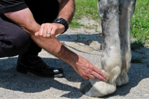
Horses developing laminitis might shift the weight off their feet twice as much as they do normally. Photo: iStock Your horse’s best chance of overcoming this hoof disease might lie in your ability to catch it early It’s a painful condition that veterinarians, farriers, and horse owners have been racking their brains about for decades. Laminitis—the separation or failure of laminae, which connect the hoof wall to the coffin bone within—can cause permanent structural changes in a horse’s foot, leading to repeated bouts of disease and lasting lameness. In severe cases the pedal (coffin) bone in the hoof rotates downward, potentially even puncturing the sole and prompting the decision to euthanize. But get this: Watchful handlers can actually detect signs of laminitis in its early stages and intervene before the condition becomes debilitating. “Everyone talks about laminitis being a lameness issue, but we know that horses start to get damage at a microscopic level before they show any lameness,” says Andrew van Eps, BVSc, PhD, MACVSc, Dipl. ACVIM, senior lecturer and specialist in equine medicine at The University of Queensland Equine Hospital, in Gatton, Australia. Therefore, keeping an eye out for minute changes in your horse’s health is key to maximizing his likelihood of recovery, says Tom Ryan, FWCF, a researcher and farrier based in Bedfordshire, U.K. “You have to be proactively thinking ahead,” he says. To help you catch this devastating hoof disease while your horse still has a chance to avoid suffering its consequences, our sources have helped us come up with a list of 10 early warning signs. Regardless of the type of case (-supporting-limb, systemic inflammatory response syndrome, or endocrine disease-related), these red flags could indicate laminitis is setting in—even before you see any signs of lameness. So alert your veterinarian as soon as possible if you detect one or more of the following:
  1. A strong/bounding digital pulse 
Slide your hand down the side of your horse’s lower limb where the digital artery runs through the groove between the flexor tendons and the suspensory ligament. Where the artery continues down the back of the fetlock you should be able to feel his pulse. Normally, the pulse should be faint or even seem absent, says Ryan. But in laminitic horses it will be stronger and is often referred to as “bounding.” How do you know what’s strong or bounding? “You kind of have to feel to know,” he says. Ideally, you should get to know what’s normal for your horse’s digital pulse. A strong digital pulse can indicate other foot pain, as well, but a bounding digital pulse in both feet is a major clue that laminitis is to blame.
  1. A hoof that’s hot for hours
Healthy horses can have hot hooves, says van Eps, but not for long periods of time. It’s normal for horses to experience large influxes of blood into their feet periodically, which causes hoof temperature to rise. But the body regulates this heat, and it should only last a couple of hours at most—unless it’s hot outside. In other words, there’s no need to panic if your horse has been standing in a sunny field on a 90-degree day and his hooves are hot. The time to worry is when hooves reach 91.4°F (33°C) for several hours in a row and the outdoor temperature is lower than 77°F (25°C). “That’s a sign they could be getting laminitis, and that’s what we’ve seen experimentally,” van Eps says. That increased temperature, says Ryan, is the hoof’s response to the trauma within the laminar tissues. Sometimes lameness follows quickly. “We see lameness begin eight to 12 hours after that temperature increase,” van Eps says. If you don’t think you can estimate surface temperatures well with your fingers, van Eps recommends using an infrared surface temperature gauge from a hardware store.
  1. A distorted hoof shape and/or unusual rings 
Healthy hooves grow faster in the dorsal (front) part of the hoof and slower in the quarters, says Ryan, creating smooth, wide, evenly spaced growth rings across the front of the hoof wall, which can be slightly closer together at the quarters. With laminitis, that growth pattern no longer applies; because the growth at the heels is now more rapid than at the toe, the horse develops wider growth rings at the heels. Currently, says Ryan, we don’t understand why the heels grow faster than normal in laminitis. This altered pattern causes the hoof’s rings to curve upward and abnormal rings to develop on the hoof wall surface, which can precede lameness sometimes by months or years, says Donald Walsh, DVM. Walsh leads the Animal Health Foundation, in Pacific, Missouri, which funds research and education projects related to laminitis. Ryan believes abnormal hoof growth starts before all evidence of pain, causing the laminae to be traumatized when the hoof capsule changes shape around the underlying pedal bone. He’s been investigating the pain-relieving properties of cutting vertical grooves into laminitic horses’ hoof walls, with the intention of reducing the effect of the distortion. The small strip of horn removed protects the dorsal laminae by isolating it from the rapidly growing heel. “Early grooving appears to protect the laminae from the more severe effects of hoof capsule distortion,” he says, adding that research is pending on this theory. “Our experience indicates that if you can do it very early on, you can see pain relief within hours.” A strong or bounding digital pulse in both limbs is a clue that laminitis is to blame. Laminitis Pulse

Photo: Stephanie L. Church, Editor-in-Chief

  1. An increased heart rate 
“We used to think that heart rate was not a very sensitive indicator,” van Eps says. “But we found that in our clinical patients the most sensitive indicator (for pending laminitis) is probably an increase in heart rate.” Most horses maintain pretty consistent resting heart rates of 30-40 beats per minute (BPM). But van Eps has noted that heart rates of -laminitic horses tend to rise a day or so before lameness sets in. “A mild increase in heart rate of even 6 bpm can be a significant early indicator that your horse is getting uncomfortable,” he says. “A lot of people have dismissed small heart rate changes, but actually they can be very useful warning signs if the horse’s normal heart rate is known,” he adds. You can measure heart rate with a stethoscope or by feeling for the pulse under the jaw or at the pastern, as described before. Exercise, excitement, and ambient temperature can also increase heart rate, however, so keep these influences in mind when interpreting results.
  1. Too little—or too much—foot lifting
Horses rely on movement to get blood flow and the nutrients within to hoof tissues, says van Eps. But if a horse is injured in one leg, he might bear weight for too long on the opposite limb, causing a phenomenon called “supporting-limb” laminitis. “Consider putting these horses in a sling so they can take weight off that foot,” suggests Walsh. With your veterinarian’s direction you can also try to get horses with leg injuries out of the stall to move around. “Even if the horse is hopping, it’s producing enough movement to prevent laminitis from occurring.” On the flipside, a horse that picks his feet up too often might also be showing early signs of laminitis, van Eps says. “Horses normally shift their (weight between) feet about two or three times per minute,” he says. “We noticed an increase of three to five times that weight-shifting when they were developing laminitis.” If the feet get painful enough, the horse will begin to change his stance, shifting his weight back to his haunches, along with stretching his legs out in front of him in the classic laminitis pose.
  1. Apparent stretched and/or bleeding laminae 
As laminae start to stretch, they separate from the hoof wall, explains Walsh. A gap becomes visible along the white line, where the sole and hoof wall meet. This white line widening is known as “seedy toe,” and you can see evidence of this in the trimmings from a laminitic horse’s foot during a farrier visit. He adds that if you notice spots of blood in the white line when you pick up your horse’s foot, it doesn’t mean your horse has been quicked by the farrier; it means the laminae are hemorrhaging, which is a sure sign of laminitis. Owners can check trimmings each time the farrier comes and begin to recognize what’s normal and not normal for their horses—something farriers might miss, says Walsh. Laminar stretching also increases the distance between the external hoof wall and the front of the coffin bone, which can be seen on lateral radiographs (X rays), says van Eps. “That space is normally 18 mm, but it will increase as the laminae stretch,” he says. “There won’t be any rotation of the bone yet, just that lengthening that could go to 20, 21, sometimes even 22 mm.”
  1.  A shortened stride 
A laminitic horse starts shortening his stride before he begins limping, says Ryan. Stride changes are more obvious on hard surfaces, especially when turning at the walk. “Not many other conditions will make a horse lame on a circle on a hard ground at the walk in both directions,” van Eps says. Know your horse’s typical stride length at the walk so you’ll recognize when those steps get shorter. And watch for signs of pain when the horse turns at the walk; these can range from the horse pinning his ears to a reluctance to move. “A horse that looks fine on a straight line on soft ground could look very different on hard ground in a circle,” he adds.
  1. Increased insulin levels  
Insulin, a normal hormone released in the body to regulate blood sugar, activates a particular growth factor (IGF-1) in the laminae, causing them to grow. “But the laminae aren’t supposed to grow,” Walsh says. A basic insulin reading by a veterinarian should show 20 units or lower, Walsh says. If it’s over 40, you need to take action to get that insulin down before laminitis hits. In the “gray area” between 20 and 40, Walsh says he recommends performing an oral glucose test, in which the horse receives a weight-calculated dose of corn syrup that causes insulin levels to spike. In healthy horses these levels should return to normal within 60 to 90 minutes. In insulin-resistant horses they’ll stay elevated for much longer. If your horse is already experiencing a bout of laminitis, however, it’s important to wait until it has subsides to test for IR. Walsh says he sees “storms” of laminitis cases caused by high insulin in spring, when horses are turned out on sugar-rich pastures. Even so, rich spring pastures won’t affect all horses—just those genetically inclined to insulin resistance. These animals need low-sugar diets and plenty of exercise—even if they’re -laminitic—to lower their insulin levels, Walsh says.
  1. Obesity 
An obese horse is more likely to be an insulin-resistant one, say our sources. Obese horses’ feet also bear more weight, which contributes to the mechanical changes in hoof shape. “Physical weight could cause the hooves to grow abnormally,” says Ryan. The excess weight can also send conflicting messages to farriers, says Walsh. “A lot of farriers will think the horse is wearing his feet down from being overweight,” he says, when in reality, “the horse is breaking his foot down, not wearing his foot down, due to first the stretching of the laminae. Then the excess weight helps to break the foot down.” First and foremost, it’s important to recognize if your horse is fat. “Owners are generally blind to how overweight their horses are and don’t appreciate how much they’re overfeeding,” says Ryan. And, secondly, reduce the horse’s calorie intake and increase his exercise, Walsh adds.
  1. Diarrhea, infection, or inflammatory response
Systemic inflammatory responses can trigger laminitis, says Walsh. “If the horse is developing a high fever and diarrhea, then laminitis is next on the plate, and you have to be proactive in thinking about that,” he says. “These horses are engaged in massive destruction of the laminae, which occurs because of enzyme reactions in the feet due to the inflammatory response.” A good way to keep laminitis at bay in these types of cases is packing the horse’s feet in ice, Walsh says. Work with a veterinarian to keep the horse’s legs at a maximum of 41°F (5°C) all the way up to his hocks and knees, starting immediately. If you wait to do this until clinical signs of laminitis appear, you’re often too late to prevent damage. Take-Home Message Laminitis starts at a microscopic level well before actual lameness sets in. Careful horse owners can learn to recognize the subtle signs caused by microscopic changes, call their veterinarian, and take steps to prevent the laminitis from worsening. Although these microscopic changes might not be reversible, it’s possible to stop the disease before it becomes debilitating. “Laminitis is all about ameliorating or halting the progression,” van Eps says. “If it’s very early and quite mild then you can go on to have a horse with no functional disruption.” Researchers recommend getting an idea of what’s normal for each horse—how he walks and turns, how he stands, what his posture is like, and his normal hoof temperature and heart rate. “Once you do that,” van Eps says, “you’ll be well-equipped to detect what’s not normal and instigate change.” ABOUT THE AUTHOR Author Laminitis Christa Lesté-Lasserre, MA Christa Lesté-Lasserre is a freelance writer based in France. A native of Dallas, Texas, Lesté-Lasserre grew up riding Quarter Horses, Appaloosas, and Shetland Ponies. She holds a master’s degree in English, specializing in creative writing, from the University of Mississippi in Oxford and earned a bachelor’s in journalism and creative writing with a minor in sciences from Baylor University in Waco, Texas. She currently keeps her two Trakehners at home near Paris. Follow Lesté-Lasserre on Twitter @christalestelas.    

Equine Protozoal Myeloencephalitis (EPM)

Know the Signs of EPM, a Master of Disguise In addition to an altered gait, horses with EPM can have paralysis of muscles of the eyes, face, or mouth, evident by drooping eyes, ears, or lips.   EPM Horse

Photo: Noah D. Cohen, VMD, PPH, PhD, Dipl. ACVIM

Equine protozoal myeloencephalitis (EPM) is a debilitating neurologic disease that cannot be ignored. More than half of all horses in the United States—and in some areas as many as 90%—have been exposed to the disease’s causative agents. Its ability to masquerade as other health issues, such as lameness or other neurologic diseases, can make it difficult to diagnose. While Sarah Reuss, VMD, Dip. ACVIM, of Merial Veterinary Services, says some horses exposed to EPM’s causative agents will not develop the clinical disease, horses under stress are more likely to show signs. In some cases, horses can even be infected without showing signs for months or years or ever. When horses do show signs, it can go misdiagnosed because it can slowly progress and infect any portion of the central neurologic system, mimicking other conditions. Left untreated, EPM can lead to permanent damage. Reuss emphasizes the importance of early detection by horse owners and farm managers, veterinary diagnosis, and an effective treatment plan. “These are the keys to stopping the progression of the disease,” she says. “The faster treatment begins, the better the chance for the horse to recover.”   How Does a Horse Get EPM in the First Place? It starts with a protozoal parasite, most commonly Sarcocystis neurona, via the opossum after it ingests contaminated tissue from intermediate hosts such as the armadillo, skunk, or raccoon. In its infective stage, the sporocysts are passed through the opossum’s feces, which the horse comes into contact with as he grazes or eats or drinks contaminated feed or water. Once consumed, the sporocysts travel from the intestine into the bloodstream and cross the blood/brain barrier where they can cause inflammation and damage the horse’s central nervous system. Researchers now know that a second organism—Neospora hughesi—can also cause EPM, however this protozoan parasite’s life cycle is less well-understood. Early Diagnosis is Crucial “EPM can evolve slowly or present suddenly with varied signs from mild to severe,” Reuss says. There are several factors that can determine its progression and severity, including:
  • How long the horse has been infected;
  • The points in the brain or spinal cord where the sporocysts have infected; and
  • Stressful events during EPM infection.
“Lesions, inflammation, and pressure may develop in the brain, brain stem, or spinal cord,” Reuss says. “Depending on the location, they can affect how the disease presents itself and can make it tricky to diagnose.” She notes that the American Association of Equine Practitioners has listed these EPM clinical signs to watch for:
  • Incoordination, weakness, and/or abnormal gait;
  • Muscle loss on one side, usually along the topline or the hindquarters;
  • Paralysis of muscles of the eyes, face, or mouth, evident by drooping eyes, ears, or lips;
  • Loss of sensation of the face;
  • Difficulty swallowing; and
  • Head tilt with poor balance—the horse might assume a splay-footed stand or lean against stall walls for support.
She also notes that loss of coordination of the mouth, tongue, and throat area could make picking up, chewing, and swallowing feed difficult. When it’s EPM, Be Ready Veterinarians have several EPM treatment options available. The first U.S. Food and Drug Administration-approved EPM treatment is Marquis, an oral paste. EPM treatments are designed to cross the tough blood-brain barrier, reach the central nervous system and successfully kill the parasite responsible. If treated quickly and properly, horses can recover from EPM. The earlier treatment begins, the better the expected outcome.     Take-Home Message Reuss emphasizes the importance of understanding the risk and knowing the signs: “With an early EPM diagnosis and immediate, effective treatment, horses can show significant improvement, and they may return to normal activity.”